World’s first floor-moving, high-field
1.5T intraoperative MRI (iMRI) solution
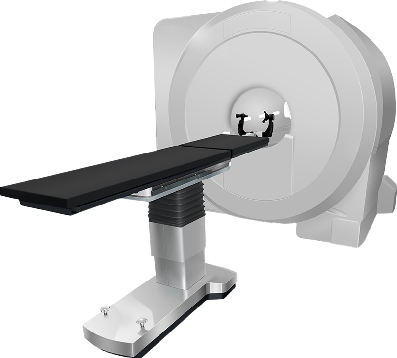
System Components
High Field 1.5T MRI System
High-field 1.5T cryogen-free MRI with 70cm bore and AI-powered image reconstruction/noise-reduction technology.
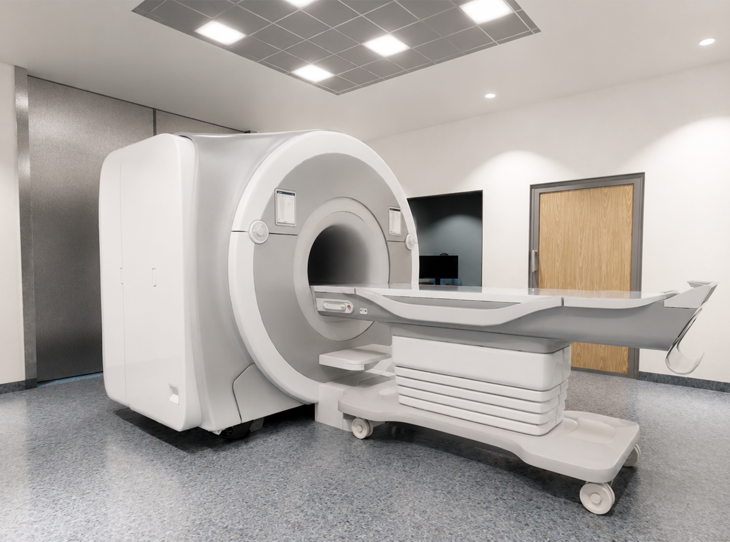
Magnet Transport System (MTS)
Magnet Transport System (MTS) mobilizes 1.5T MRI between OR and Diagnostic Room (DR) or Magnet Storage Bay (MB) with a floor-moving tracked crawler. Designed for less than 3-minute transit time with sub-millimeter positioning including anti-collision detection and safety interlock.
MR-Compatible OR Table
MR-compatible neurosurgical OR table designed for intraoperative imaging. Multi-axis adjustment including horizontal deck movement up to 200mm. Table may be removed from OR to accommodate different procedures.
MR-Compatible Cranial Stabilization System
MR-compatible, radiolucent Mayfield-style skull clamp provides rigid cranial fixation during surgery and imaging. Supports prone, supine, and lateral positioning.
Flexible 8-Channel Neurosurgical RF Coil
8-channel, two-piece flexible design (anterior/posterior arrays, four channels each) compatible with Mayfield-style fixation devices. Imaging coverage brain and upper cervical spine (to C3).
Patient Imaging Positioning System (PIPS)
Projected laser crosshair reference at region of interest for magnet isocenter positioning to guide imaging accuracy and workflow.
RF & Magnetic Room Shielding
RF/H-shielded OR suite designed to protect image integrity during intraoperative scans. Pneumatic-seal doors isolate the high-field environment when MRI not in use. Allows OR to perform as conventional operating suite when magnet is stored.
Bringing 1.5T iMRI
to the Neurosurgical OR
The Taumedis Intraoperative MRI (iMRI) solution features a ground-based platform to transport a high-field 1.5T MRI system, enabling real-time imaging during neurosurgery without moving the patient.
Floor-moving tracked crawler is designed to support up to five (5) tons, while distributing magnet load over the entire track area. This system aims to reduce infrastructure requirements for intraoperative MRI, and thus make iMRI more accessible to more neurosurgical programs.
Magnet movement can be controlled in two ways:
Hand-Held Pendant
Controls magnet position to and from the OR.
Magnet Mover Software Console
Controls and monitors magnet position.
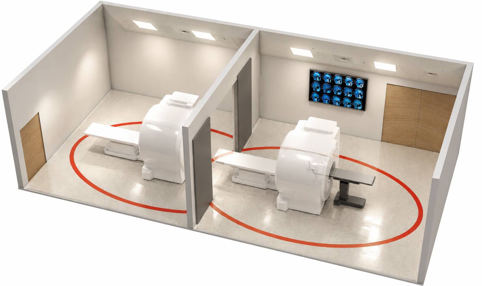
Ground-Based iMRI Transport
Floor-moving Magnet Transport System (MTS) mobilizes a 1.5T MRI, designed to follow a pre-defined optical path with sub-millimeter positioning accuracy. MRI transitions between adjacent Diagnostic Room (DR) or Magnet Storage Bay (MB) to Operating Room while patient remains stable.
Following intraoperative scans the MRI is returned to home position, either resuming diagnostic use (DR configuration) or stored until the next case (MB configuration).
See below for more details on iMRI floor plan options.
Diagnostic Imaging or Standby
Magnet travels between adjacent Diagnostic Room (DR) or Magnet Storage Bay (MB) to operating room guided via optical reference path. Transport can be controlled from OR with handheld pendant or console interface. Safety features include Anti-Collision System (ACS) and real-time transport monitoring.
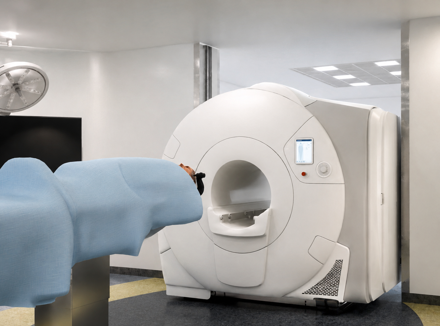
Moving Into OR for Intraoperative Scans
MRI guided into position in the OR for intraoperative scans via foor-based Magnet Transport System (MTS) while patient remains stable. MR-Compatible neurosurgical table, MR-Compatible Cranial Stabilization System, and RF-shielded room serve to protect image quality from artifacts.
Room Configurations
The Taumedis system is designed to be configured in two broad formats:
Multi-purpose Operating/Diagnostic Room (OR/DR)
Space-saving OR with Magnet Storage Bay (OR/MB)
When not in use during surgery, the MRI is housed in the adjacent DR for conventional diagnostic imaging, or stored in its bay to maximize OR space between cases.
Shielding for Image Quality and Safety
RF (radiofrequency) and H (magnetic) shielding help protect image integrity during scans, while containing the always-active magnetic field between rooms. As such, the OR remains magnetically “quiet” when the MRI is located in the adjacent DR or MB. An RF/H shielded sliding door separates the adjacent rooms.
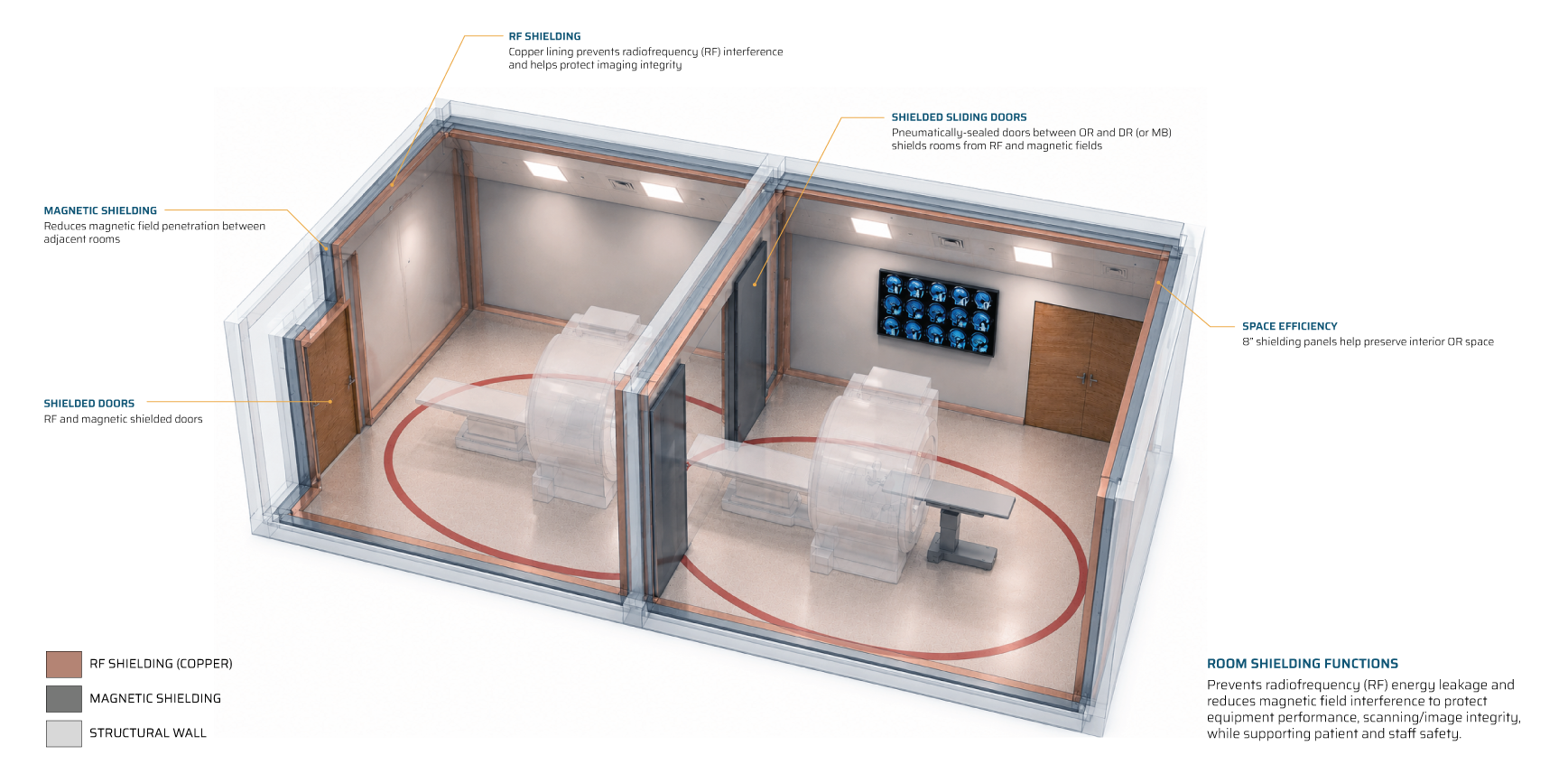
Other floor plans can be considered based on available space and clinical requirements.
Contact us to discuss configuration options
Multi-Purpose Configuration (OR/DR)
Combines adjacent Diagnostic Room (DR) for use when OR is not active.
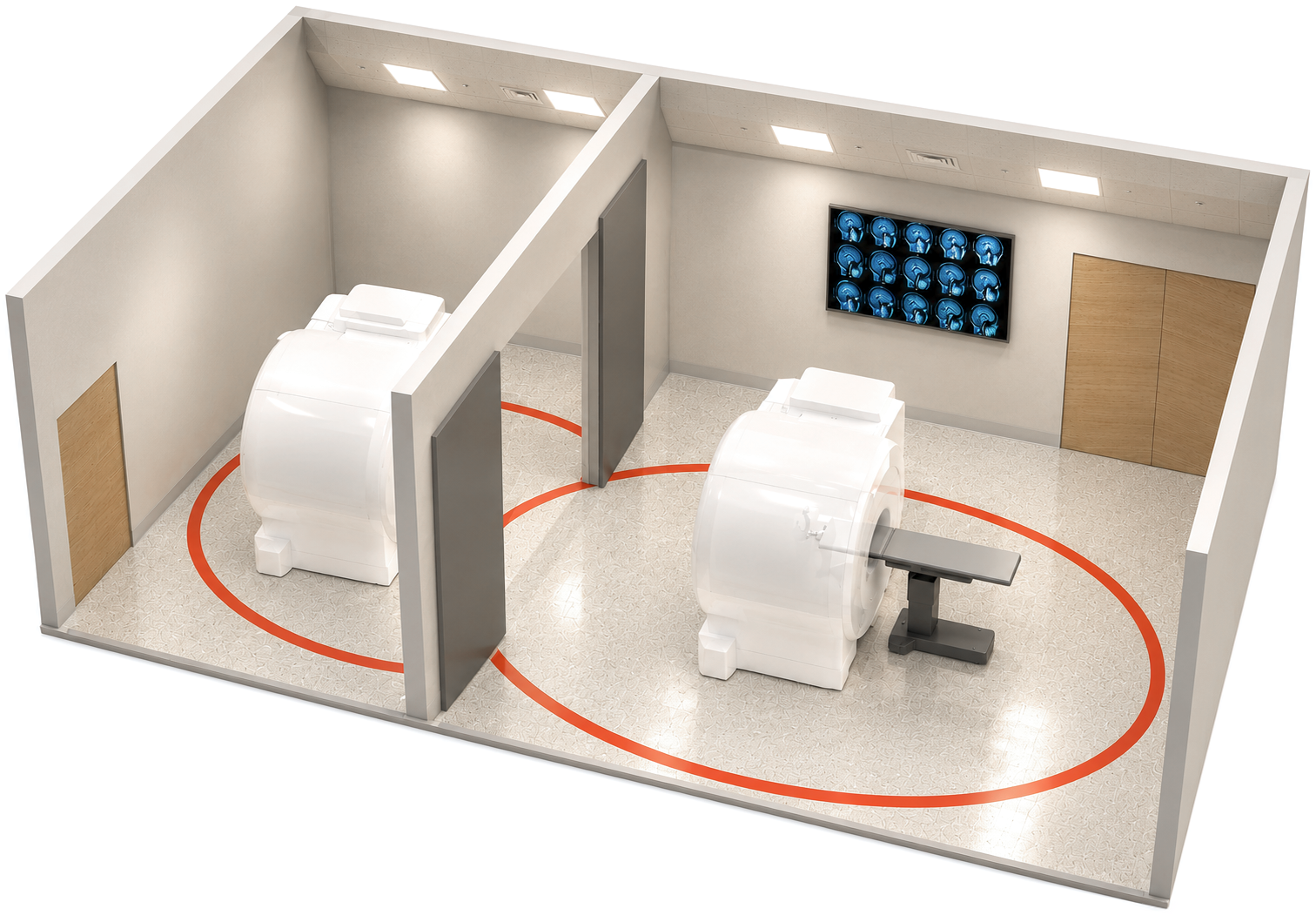
Dedicated Intraoperative OR Configuration (OR/MB)
Magnet Storage Bay (MB) houses MRI when OR is not active.
This device is currently under development, not yet authorized for sale by FDA or Health Canada. Not for clinical use or distribution within USA or Canada. Specifications are preliminary and subject to change prior to regulatory clearance.
Intraoperative MRI (iMRI)
Frequently Asked Questions
What is Intraoperative MRI (iMRI) and how is it used in Neurosurgery?
Intraoperative MRI (iMRI) is the use of Magnetic Resonance Imaging during a surgical procedure. It enables MRI images to be acquired at select points throughout the procedure to support surgical decision-making, thereby reducing reliance on post-operative imaging as the first opportunity to evaluate surgical results.
Published neurosurgical studies1,2 have associated intraoperative MRI with improved extent of resection in selected brain tumor procedures and have examined its potential to reduce early repeat surgery by identifying residual tumor before the procedure is completed.
iMRI systems can be configured using either a moving-magnet or patient-moving workflow and are commonly integrated into neurosurgical operating environments designed for MRI safety and compatibility.
1 Senft, C., et al (2011). Intraoperative MRI guidance and extent of resection in glioma surgery: a randomised, controlled trial. The Lancet Oncology, 12, 997–1003.
2 Berkmann, S., et al (2014). Intraoperative high-field MRI for transsphenoidal reoperations of nonfunctioning pituitary adenoma. Journal of Neurosurgery, 121(5), 1166–1175.
Is this a High-Field 1.5T Intraoperative MRI (iMRI) System?
Yes. The Taumedis iMRI solution is designed to mobilize a high-field 1.5T intraoperative MRI via a ground-based tracked Magnet Transport System (MTS).
For intraoperative MRI applications, magnet field strength and system mobility are separate. Some iMRI solutions utilize low field strength (ie. <1T) magnets, which may have limitations with signal-to-noise ratio (SNR), image resolution, scan time, and/or available imaging protocols. For this reason, field strength is commonly evaluated as part of the broader iMRI planning process—which is why the Taumedis iMRI system integrates a high-field strength 1.5T magnet.
Our ground-based moving-magnet system is designed to transport the high-field 1.5T scanner into position in the OR for intraoperative imaging without moving the patient.
What is a Magnet-Moving vs Patient-Moving Intraoperative MRI (iMRI)?
The Taumedis intraoperative MRI (iMRI) system is a magnet-moving solution, designed to transport the high-field 1.5T MRI directly to the patient in the OR as they remain stable for imaging mid-procedure.
In a patient-moving iMRI system, the patient is moved from the operating room to a nearby MRI scanner. Workflows require patient transport, introducing additional considerations for sterility, anesthesia management, monitoring equipment, patient positioning, and workflow coordination.
In a magnet-moving iMRI system—as with Taumedis—the scanner moves into the OR while the patient remains stable; this approach can simplify workflow by eliminating intraoperative patient transport and associated logistics.
Each approach involves planning considerations. Magnet-moving systems focus on magnet transport, OR integration, movement control, proper shielding, and MRI access in the surgical suite. Patient-moving systems focus on patient transfer, room/scanner access, transport workflow, and maintaining surgical setup during movement.
How does your ground-based moving-magnet Intraoperative MRI (iMRI) system work?
Simply put, a moving-magnet iMRI system brings the MRI scanner to the patient in the OR, rather than transporting the patient mid-procedure to the scanner. The magnet moves into the operating room when imaging is needed, then returns to its origin when imaging is complete. The patient remains stable on an MR-compatible surgical table while the scanner is brought into imaging position.
This approach is built around controlled magnet movement, a defined travel path, MRI safety procedures, and integration with the surgical environment/workflow.
Taumedis’ iMRI solution integrates a ground-based Magnet Transport System (MTS) to mobilize a high-field 1.5T MRI from an adjacent Diagnostic Room (DR) or Magnet Storage Bay (MB) into the operating theater. The tracked MTS is designed to distribute the system weight over a broad area, follows a pre-defined path between locations, and can provide sub-millimeter positioning accuracy.
An overhead laser crosshair (Patient Imaging Positioning System) helps align the region of interest with the magnet’s isocenter. Magnet movement can be managed from within the operating room or from an adjacent control room. Multiple safety systems track magnet movement from start to finish.
To see the Taumedis ground-based iMRI system in action, please see our video above and on our YouTube page.
What is a Diagnostic Room vs. Magnet Storage Bay for an Intraoperative MRI (iMRI) suite?
Intraoperative MRI (iMRI) systems move the magnet between adjacent spaces, to and from the operating room. A Diagnostic Room (DR) and Magnet Storage Bay (MB) describe where the MRI scanner is located when it is not being used during an active procedure in the OR. The preferred iMRI configuration depends on (but is not limited to) space, construction goals, surgical volume, diagnostic imaging needs, staffing, and room utilization.
The Taumedis iMRI solution can accommodate these configurations among others.
- Diagnostic Room Conventional MRI room adjacent to OR, used for diagnostic scanning between surgical cases.
- Magnet Storage Bay Dedicated space for MRI adjacent to the OR. Primarily planned around intraoperative use and stored in the bay (albeit still “live”) between surgical cases.
In either configuration, the Taumedis iMRI solution includes room shielding against RF (radiofrequency) and H (magnetic) interference. The former helps protect image integrity, while the latter contains the always-active magnetic field between rooms. As such, the OR remains magnetically “quiet” when the MRI is located in the adjacent DR or MB. An RF/H shielded sliding door separates the adjacent rooms.
To see the Taumedis iMRI system in an OR/DR configuration, please see our video above and on our YouTube page.
How does intraoperative MRI (iMRI) impact surgical workflow?
Intraoperative MRI (iMRI) procedures include MRI safety preparation, equipment positioning, image acquisition, and return to the surgical field. The downstream value of this time investment is how imaging during surgery helps the team assess for residual tissue before the operation is completed; published evidence has associated iMRI with improved extent of resection and reductions in repeat surgery when residual tissue would otherwise be identified after the procedure.1,2 A recent paper noted improved iMRI efficiencies with increased use, and a decreasing trend in the time taken per case.3
Our Clinical Specialist will work closely with your team to streamline and refine your iMRI implementation, consistent with local policies and procedures.
Workflow impact is ultimately a function of system integration (ie. moving the magnet vs moving the patient), site protocols, and the types/number of scans required. For clinicians evaluating iMRI, workflow should consider how associated imaging steps may be offset by the ability to assess surgical progress before the patient leaves the OR, and how this relates to re-surgery rates.
1 Senft, C., et al (2011). Intraoperative MRI guidance and extent of resection in glioma surgery: a randomised, controlled trial. The Lancet Oncology, 12, 997–1003.
2 Berkmann, S., et al (2014). Intraoperative high-field MRI for transsphenoidal reoperations of nonfunctioning pituitary adenoma. Journal of Neurosurgery, 121(5), 1166–1175.
3 Madani, D. et al (2025). Comparing the Rates of Further Resection After Intraoperative MRI Visualisation of Residual Tumour Between Brain Tumour Subtypes: A 17-Year Single-Centre Experience. Brain Sciences, 15(1).
What factors influence the cost of an Intraoperative MRI (iMRI) system?
The cost of intraoperative MRI is shaped by the full project, not the scanner alone. Hospitals typically consider acquisition cost, construction, shielding, room design, MR-compatible equipment, surgical table integration, service, training, staffing, and workflow implementation.
Cost drivers can vary by iMRI implementation. Moving-magnet systems may require additional infrastructure and structural planning. While patient-moving iMRI systems may reduce magnet transport requirements they also impact planning around patient transfer, anesthesia, sterility, and/or adjacent-room access. A high-field ground-based moving-magnet system brings the MRI to the patient and can mitigate challenges with extensive infrastructure or access.
Hospitals should also consider indirect costs, including OR downtime during renovation, installation complexity, construction phasing, and whether the MRI space can support diagnostic imaging when surgery is not active. Not all systems are created equally, and allocated clinical space and requirements vary.